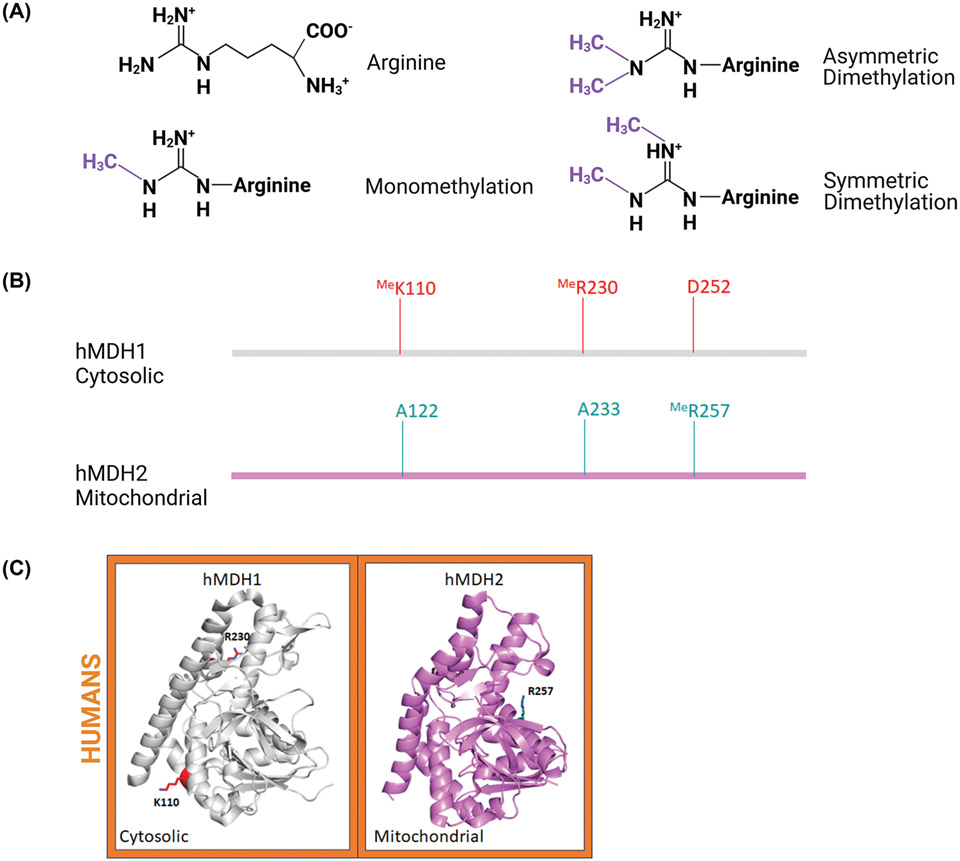
Figure 5. Sequence and structural comparison of methylated (Me) residues identified on human MDH proteins.
(A) Chemical structures of known arginine methylations. (B) Methylated residues represented on the linear sequence of human MDH isoforms. (C) Crystal structure of monomeric human MDH1 (PDB: 7rm9; UniProtID: P40925) and MDH2 (PDB: 4wlo; UniProtID: P40926) isoforms with methylated residues. These structures are colored as described in Figure 2. Three-dimensional structures were generated using PyMOL and the figure was constructed using BioRender.com.

