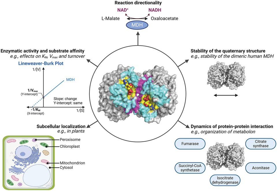
Figure 6. Potential roles of PTMs on MDH.
At the center, the three-dimensional structure of the porcine cytoplasmic MDH dimer is represented (PDB: 5mdh). The active site is highlighted in yellow, the dimer interface in magenta, and the citrate synthase surface for the metabolon complex in cyan. The NAD+ ligand is shown in yellow spheres. Properties that can be modulated by PTMs, including defining the directionality of the reaction, altering enzymatic characteristics (e.g. Km and Vmax), defining the subcellular localization, regulating the stability of the quaternary structure and protein–protein interactions protrude around the circle. This figure was generated using PyMOL and BioRender.com.

