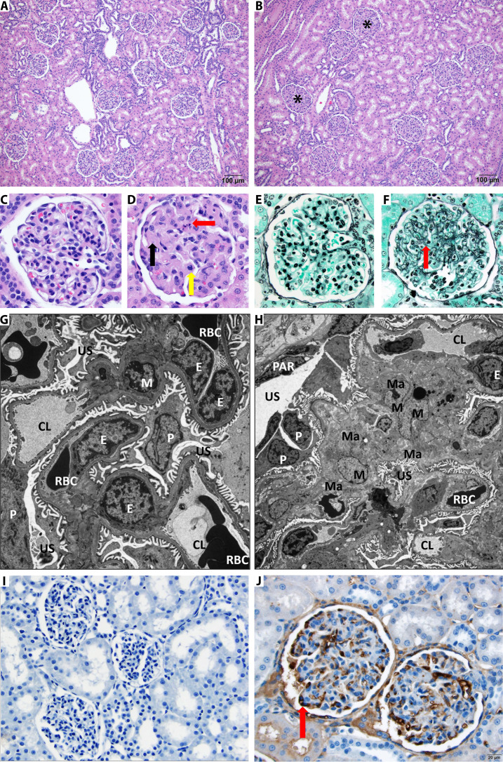
Fig. 4. Multiple dose administration of AM1-15 to cynomolgus monkeys causes unique renal toxicity.
Hematoxylin and eosin–stained kidney from control (A) and treated cynomolgus monkey (B). There is diffuse thickening of glomerular matrix in the AM1-15–treated kidney (asterisk). Scale bar, 100 μm. At high magnification as compared to control monkey glomeruli (C), the AM1-15–treated glomeruli (D) has global thickening of glomerular matrix by eosinophilic material (black arrow), and occasional degenerate cells (red arrow) and mitotic figures (yellow arrow) are also present. Periodic acid methenamine stain revealed an increased mesangial matrix component (red arrow) in the AM1-15–treated glomeruli (F) as compared to control monkey glomeruli (E). Electron microscopy revealed enlarged mesangial cell with increased cytoplasm (hypertrophy) along with moderate increase in extracellular mesangial matrix deposition in the AM1-15–treated glomeruli (H). The rest of the glomerular components, namely, endothelial cells, podocytes, and filtration apparatus, was within normal limit as compared to control glomeruli (G). M, mesangial cell; Ma, mesangial matrix; E, endothelial cells; P, podocyte; PAR, parietal epithelium; US, urinary space; CL, capillary space; RBC, red blood cell. Magnification, ×6000 (G) and ×3000 (H). (I and J) Anti–BCL-XL drug-linker–specific monoclonal antibody was synthesized, and IHC was performed to visualize the distribution of drug-linker. Moderate-to-strong immunoreactivity was observed in glomerular endothelial cells (red arrow) in a cynomolgus monkey administered AM1-15 (J) (scale bar, 20 μm) as compared to control (I).

