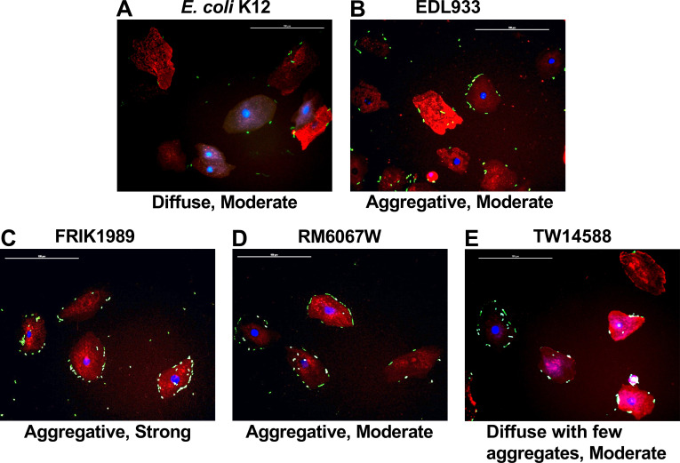
Fig 3.
Adherence patterns of E. coli K12 and E. coli O157:H7 isolates on RSE cells. E. coli isolates (K12, EDL933, TW14588, FRIK1989, and TW14588) were incubated with RSE cells at an MOI of 10:1 for 4 h with agitation followed by assessment of cell attachment via immunofluorescence staining and co-localization. Representative immunofluorescent images with RSE cells and E. coli are shown at 400× magnification. Adherence patterns on RSE cells were qualitatively recorded as diffuse or aggregative, with strong or moderate qualifiers as described in the Materials and Methods. E. coli were labeled with FITC (green) conjugated antibodies. RSE cell cytokeratins were labeled with Alexa Flour 594 (red) antibodies. RSE nuclei were stained with DAPI (blue). Scale bar represents 100 µm. Abbreviations: recto-anal junction squamous epithelial (RSE), multiplicity of infection (MOI).

