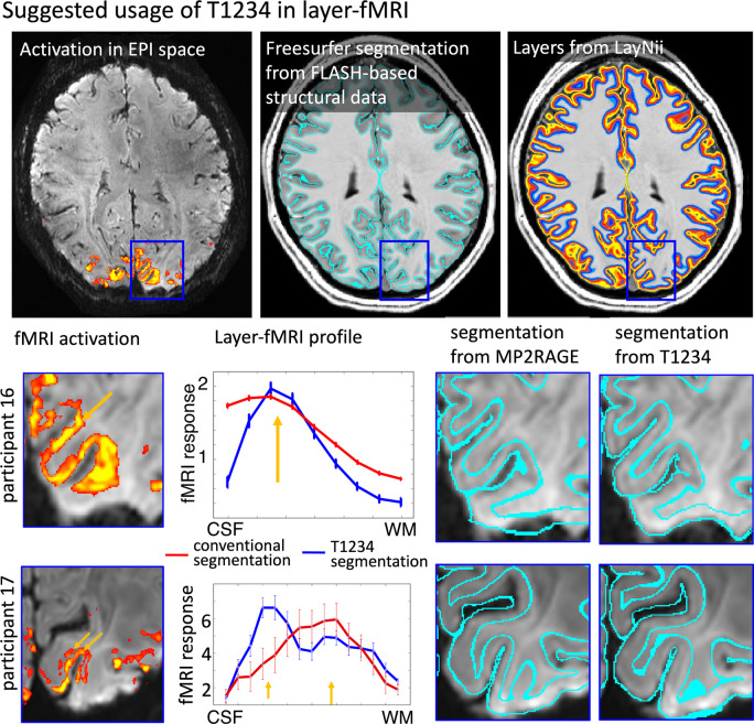
Fig. 5:
Proposed usage of T1234 for high resolution layer-fMRI applications Conventional layer-fMRI activation mapping typically uses FLASH-based structural reference data. These structural reference data are segmented in software tools like FreeSurfer to estimate layers for the extraction of activation scores across cortical depth. However, if the alignment between tissue-type borders (as derived from structural reference data) does not perfectly match the locations of functional activation data, the resulting layer profiles are compromised.
The panels for participants 16 and 17 illustrate two challenges of the conventional layer-mapping approach. The orange arrows point to layer-specific activation patterns that follow the cortical ribbon at unique cortical depths. However, when these signals are pooled from compromised segmentation borders of conventional structural reference data, the depth-dependent peaks are blurred. When segmentation borders have higher spatial precision from T1234 data, the peaks of depth-dependent activations become visible in layer profiles.

