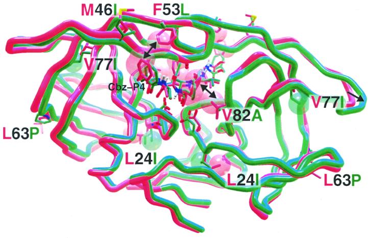
FIG. 3.
Energy-minimized model of R8 wild-type (red) protease overlaid with the TL-3 resistant 6x mutant (green) protease. TL-3 is bound to R8 and mutant proteases. Depicted are the protein backbones with mutated residues at numbered positions. The double-headed arrows indicate loss of both the TL-3 P1 interaction (P1′ not shown) with V82 upon mutation to the smaller A and the P4-Cbz interaction with the F53 upon mutation to the smaller L. The single-headed arrow points to distended flaps. The van der Waals radii of the L24I and V77I mutant side chains, resulting in rearrangement of local packing, are also shown as red (wild-type) or green (mutant) spheres, respectively.

