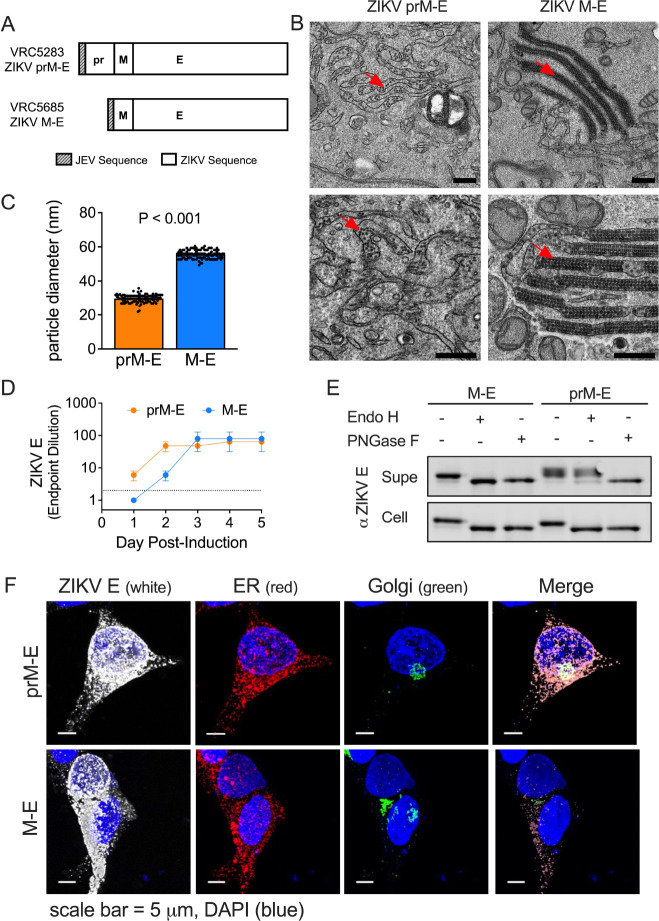
Fig. 1. pr-independent biogenesis of ZIKV subviral particles.
(A) DNA plasmids VRC5283 and VRC5685. (B) Transmission electron microscopy images of ZIKV SVPs in tetracycline-induced CHO cells 2 days post-induction. Red arrows indicate SVPs. Scale bars represent 0.5 microns. (C) Average SVP diameter calculated from 101 and 105 individual particles from prM-E and M-E images, respectively. Error bars indicate the standard error. (D) ZIKV E protein ELISA endpoint dilutions for supernatants sampled from tetracycline-induced CHO cell lines incubated at 37°C. Data is the average of two independent experiments. Error bars indicate the range. Samples that were not positive at an initial 1:2 dilution (dotted line) were assigned an endpoint dilution of 1. (E) M-E and prM-E ZIKV SVPs collected from the supernatant (Supe) or from inside transfected 293T cells (Cell) were left untreated or incubated with Endo H or PNGase F glycosidases, followed by SDS-PAGE and Western blotting with a ZIKV E protein-specific mAb. (F) Confocal microscopy of 293T cells transfected with ZIKV prM-E or M-E expressing plasmids. Transfected cells were fixed, and stained with ER, Golgi, and ZIKV E protein-specific antibodies. Scale bars represent micrometers.

