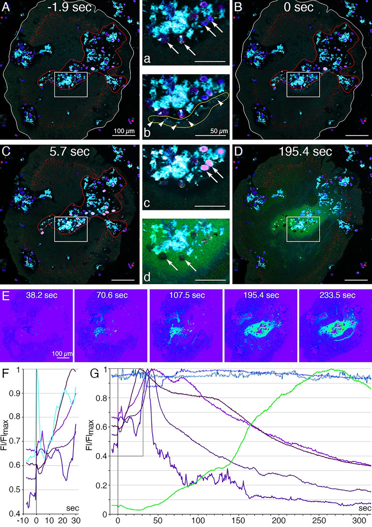
Fig 8. Secretory behaviors of Trichoplax associated with external digestion of algae.
(A-E) Confocal images of a Trichoplax feeding on R. salina algae; (F, G) normalized fluorescence intensity measured in selected regions. A starved animal was transferred to a cover glass chamber containing algae in seawater with a lipophilic dye (LipidTOX, red) that stains lipophil cell granules; FM1–43 (cyan), a membrane dye used here to visualize the secreted contents of lipophil cell granules; and a fluorescent indicator for trypsin activity (BZiPAR, green). The insets numbered with lowercase letters are enlarged view of rectangular region on a respective image numbered with an uppercase letter. (A, a) At t=−1.9 sec, the animal had ceased moving in a region containing algae (blue, phycoerythrin autofluorescence, arrows) and debris (cyan) representing algae remnants from an earlier feeding episode. The animal body is outlined white, and the feeding pocket is outlined red. (B, b) At t=0 sec, lipophil cell granule secretion was evident in the feeding pocket due to the sudden appearance of small (<5 μm) diffuse clouds and bright particles (arrowheads on b) of FM1–43-stained material. (C, c) At 5.7 sec, some algae near sites of lipophil granule secretion swelled, lysed, and became intensely stained with LipidTOX and FM1–43 (pink in merged, arrows in c). Other algae (blue) remained intact and were not stained with LipidTOX or FM1–43. (D, d) By 195.4 sec, diffuse BZiPAR fluorescence (green) filled the feeding pocket. The lysed algae no longer were visible, but the intact algae remained (arrows in d). (E) Intensity encoded BZiPAR fluorescence images show increasing trypsin activity between 70.6 and 233.5 sec. (F) Details of first events in feeding: Lipophil granules (cyan; the fluorescence profile obtained for the area outlined yellow in b) were secreted approximately synchronously at 0 sec and this was followed by a rapid increase of fluorescence in nearby algae (four fluorescence profiles obtained for four pink algal cells in c; different shades of magenta). (G) Evidence of digestion: algae affected by lipophil granules (different shades of magenta) burst and released their content. Secretion of trypsin, as indicated by BZiPAR fluorescence (green; fluorescence profile obtained for the region outlined red in B), began 40–50 sec after lipophil discharge and was associated with a decline of fluorescence intensity of the lysed algal cells. Those algae not affected by lipophil granules remained constant in intensity throughout the feeding episode (two fluorescence profiles obtained for two individual algal cells; different shades of blue). Scale bars 100 μm.

