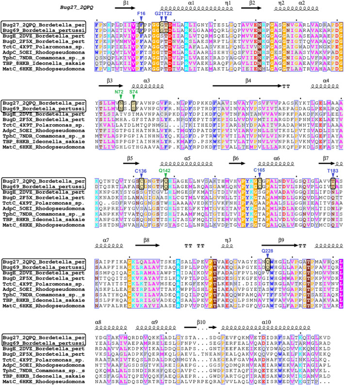
Fig. 5.

Structure‐based sequence alignment of TctC/Bug proteins with known ligand and structure. Secondary structure elements from Bug27 (PDB: 2qpq) are represented by spirals (α and 310 helices) and arrows (β strands). The 310 helix is labeled η. Partially conserved residues (numbered by Bug27 structure) are shown on a gray background, and universally conserved residues are highlighted by a solid background color. Triangles indicate residues identified in the quinolinic acid‐binding pocket, as also shown in Fig. 4. Highlighted boxes mark the unique conservation of such residues among the Bug27‐Bug69 pair. Ligands (in brackets) of TctC proteins used are as follows: Bordetella pertussis BugE (glutamate); B. pertussis BugD (aspartate); Polaromonas TctC Bpro_3516 (unknown); Rhodopseudomonas palustris AdpC (oxoadipate); Comamonas sp. TphC (terephthalate); Ideonella sakaiensis TBP ISF6_0226 (terephthalate); Rhodopseudomonas palustris MatC Rpa3494 (malate).
