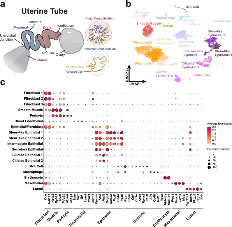
Fig. 1. Census of cell types of the mouse uterine tube.
a Diagram of a partially uncoiled mouse uterine tube. The proximal region contains few ciliated cells and extends from the intratubal junction to the ampulla. The distal region consists of the ampulla and the infundibulum where ciliated cells are abundant. b UMAP visualization of the cell types identified in a pool of 16,583 distal cells from 62 uterine tubes from which high quality sequence data was obtained. c Dot plot representation of genes associated with various tissue types to validate cell type identification. Source data are provided as a Source Data file. a was in part created with BioRender.com released under a Creative Commons Attribution-NonCommercial-NoDerivs license.

