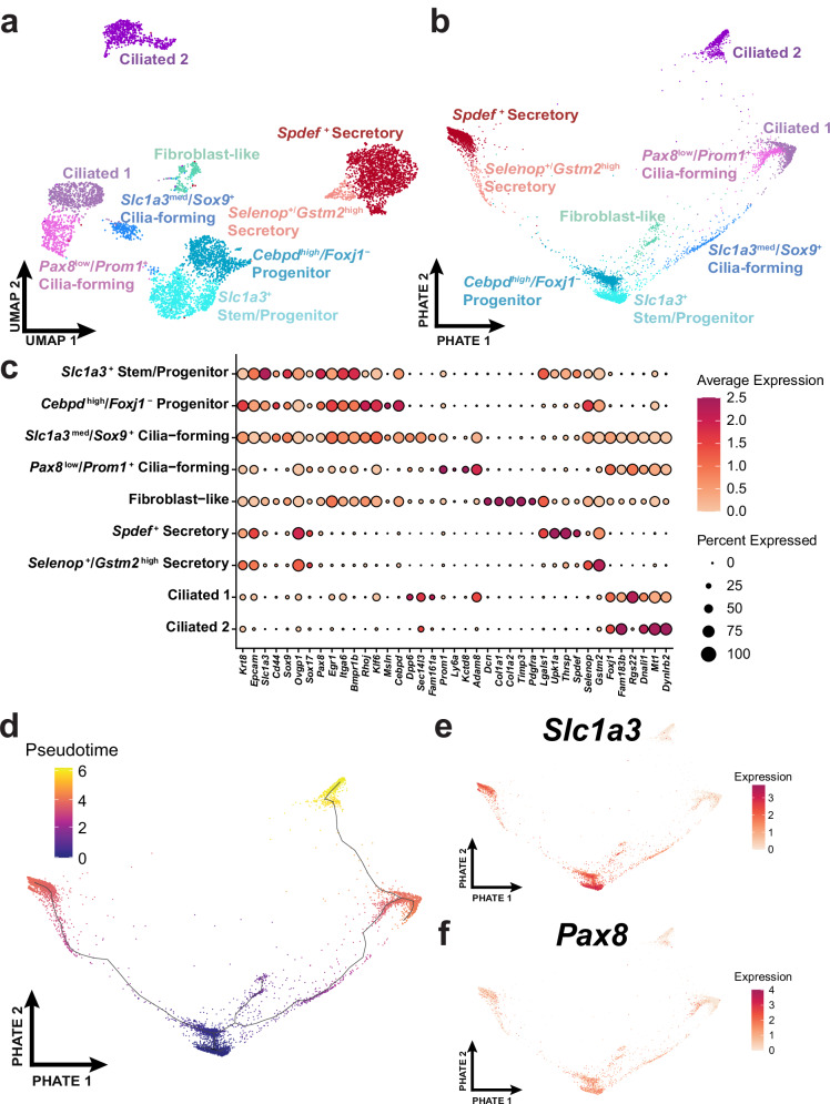
Fig. 2. Characterization of distal epithelial cell states.
a Distal epithelial cells (n = 6273) from 62 uterine tubes were identified by their Epcam and Krt8 expression and the subset was represented within the UMAP embedding. b A differentiation trajectory among epithelial cells visualized through the PHATE dimensional reduction technique. c Dot plot reflecting highly expressed, specific markers of each identified epithelial cell cluster. d Monocle3 pseudotime analyses calculated over the PHATE embedding. The expression of Slc1a3 (e) and Pax8 (f) visualized over the PHATE embedding. Source data are provided as a Source Data file.

