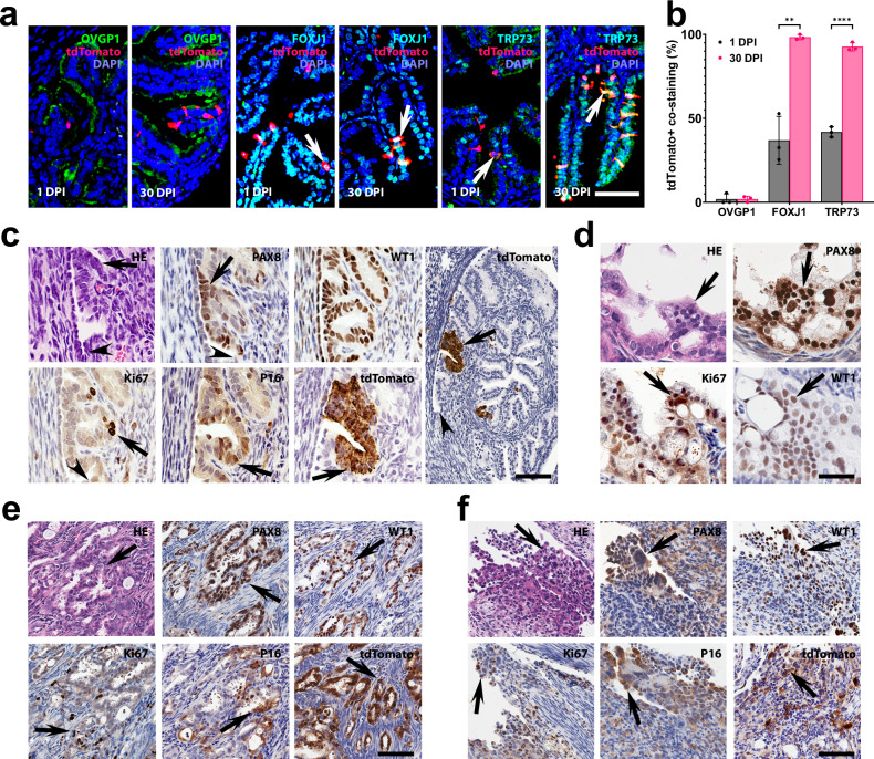
Fig. 7. Transitional pre-ciliated Krt5+ cells are highly susceptible to malignant transformation.
a Detection of ciliation FOXJ1 (green) and TRP73 (green) markers but not secretory marker OVGP1 (green) in Krt5+ (tdTomato) cells and their progeny (arrows) in the distal tubal epithelium 1 and 30 days post-induction (DPI) with tamoxifen in Krt5-CreERT Ai9 mice. b Quantification of Krt5+ cells co-expressing tdTomato with OVGP1, FOXJ1 or TRP73 1 and 30 DPI. **P = 0.0017, ****P < 0.0001, two-tailed unpaired t-tests. Data are presented as mean values ± SD. Biological replicates n = 3 in each group. Source data are provided as a Source Data file. c–f Neoplastic lesions in Krt5-CreERT Trp53loxP/loxP Rb1loxP/loxP Ai9 mice. Early dysplastic lesions (arrows) with mild cellular atypia (c) and more pronounced atypical features, loss of cell polarity, and high proliferative index typical for STICs (d). Arrowheads in (c), TE-mesothelial junctions. Advanced neoplastic lesions (arrows) with stromal invasion (e) and peritoneal spreading (f, arrows). Hematoxylin and Eosin (HE) staining and immunostainings for PAX8, Wilms tumor 1 (WT1), Ki67, p16, and tdTomato (brown color) by Elite ABC method with hematoxylin counterstaining. Scale bar 50 µm (a, c, except for right image, and d) and 100 µm (c), right image, (e, f). Biological replicates n = 5 (c), n = 8 (d), n = 4 (e) and n = 3 (f).

