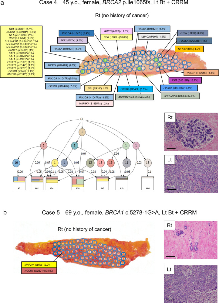
Fig. 5. Mutational mapping and phylogenetic tree analysis following contralateral risk-reducing mastectomy.
a Case 4 was a 45-year-old woman with BRCA2 p.I1065fs who had left breast cancer, underwent total mastectomy of left breast, and underwent risk-reducing mastectomy of the right breast. and case 5 was a 69-year-old woman with BRCA1 c.5278-1 G > A. Although no cancer was identified in the right breast, 14 specimens from 68 samples had mutations in PIK3CA, ARHGAP35, and PIK3R1, and the clonality score was 2.29. Phylogenetic tree analysis of 7 samples (green highlighted) using WES data are shown. The representative microscopic images of breast tissue with HE staining. No pathological finding was observed in the right breast, while the tumor in the left breast was invasive ductal carcinoma. Magnification, ×100. Scale bars represent 200 μm. b Case 5 was a 69-year-old woman with BRCA1 c.5278-1 G > A who had left breast cancer, underwent total mastectomy of left breast, and underwent risk-reducing mastectomy of the right breast. In contrast to case 4, all 68 samples from case 5 had no mutation in the 25 genes. The representative microscopic images of breast tissues with HE staining are shown in the right. Extensive hyalinization and ductal and lobular atrophy were observed in the right breast. The tumor in the left breast was invasive ductal carcinoma. Magnification, ×100. Scale bars represent 200 μm. Lt left, Rt right, GL germline.

