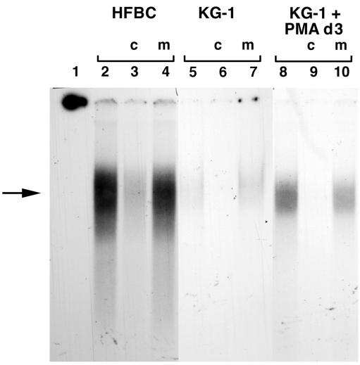
FIG. 4.
Competitive gel shift analysis of the binding of nuclear proteins from HFBC (lanes 2 to 4), untreated KG-1 (lanes 5 to 7), and PMA-treated KG-1 (lanes 8 to 10) cells to a radiolabeled oligonucleotide containing an NF-1 binding site. Competitors were either unlabeled cold homologous oligonucleotide (c) or unlabeled mutant oligonucleotide (m). Lane 1 contains the probe without any added nuclear extract and shows migration off the gel. The arrow indicates the position of a specific gel-shifted band present in HFBC and induced in KG-1 cell extracts by PMA treatment, compared to the band from extracts from untreated KG-1 cells.

