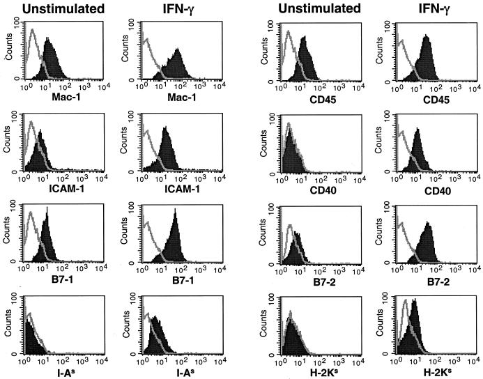
FIG. 1.
Expression of APC surface markers on unstimulated and IFN-γ-treated SJL microglia. Microglia cultures were unstimulated or stimulated with IFN-γ for 24 h. The microglia were stained with antibodies for Mac-1 (CD11b), CD45, ICAM-1 (CD54), CD40, B7-1 (CD80), B7-2 (CD86), MHC class II (I-AS), and MHC class I (H-2KS). Surface expression was then analyzed by flow cytometry, with the single line in each histogram representing the isotype antibody control and the solid peak representing the surface marker staining listed on the x axis. The flow plots shown represent all of the cells derived from each of the various culture conditions. These results are representative of four separate experiments.

