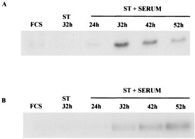
FIG. 4.
Cyclin B and cyclin B-associated kinase levels of arrested cells. (A) Confluent HDFs were infected with Ad-ST at 20 PFU/cell and then maintained in 10% FCS after infection. Extracts were prepared at 24, 32, 42, and 52 h postinfection, and levels of cyclin B were compared to those found in controls (Ad-ST alone, FCS alone) by Western blot analysis. (B) The same extracts were used for immunoprecipitation-kinase assays in which histone H1 was the substrate for cyclin B1-associated kinase activity.

