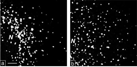
FIG. 4.
High-contrast black and white prints of maximal-intensity projections. (a) PV-infected cell stained for Sec13 and 2B. The irregularly sized colocalization signal is found concentrated in a perinuclear region. (b) Noninfected cell stained for Sec13 and Sec31. The uniform signal is rather evenly distributed through the cytoplasm. N, nucleus. Bar, 2 μm.

