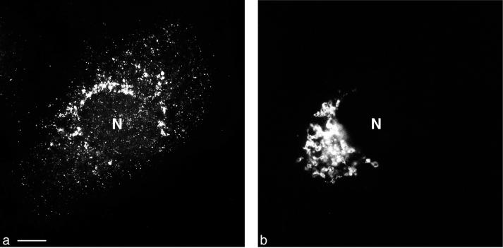
FIG. 6.
Conventional double fluorescence microscopy of a PV-infected cell. (a) Staining with FITC-tagged anti-2B MAb, enhanced with a secondary anti-FITC-Alexa 488 Ab. (b) Staining for giantin (cis-medial Golgi compartment). Distribution of the two signals in the cell is entirely different. Bar, 10 μm.

