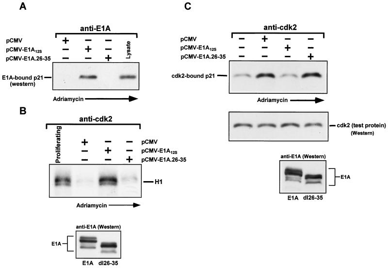
FIG. 3.
E1A expressed in doxorubicin-treated p21+/+ cells associates with p21 and reduces the amount of p21 in association with cyclin-Cdk2 complexes, thereby restoring their activity. (A) p21+/+ cells were transfected in parallel with pCMV-E1A12S, pCMV-E1A.26-35, or the control plasmid pCMV. Immediately after, doxorubicin was added to the cultures, and at 36 h posttransfection, the cells were collected and whole-cell extract was prepared. Normalized extracts were immunoprecipitated with anti-E1A, and immune complexes, along with lysate (to mark p21), were then subjected to Western blot analysis and enhanced chemiluminescence, using anti-p21 as a probe. (B) Cdk2-associated kinase activity in transfected p21+/+ cells with doxorubicin treatment for 36 h was determined as for Fig. 2. In the lower blot, the extracts used for panels A and B were subjected to Western blot analysis using anti-E1A as a probe. (C) Normalized extracts from transfected or untransfected p21+/+ cells with or without doxorubicin treatment for 36 h were immunoprecipitated with anti-Cdk2. The immune complexes were then examined for the presence of p21 by Western blot analysis using anti-p21 as a probe. The membrane was also probed with anti-Cdk2 (middle blot) to assure equal loading of the immunoprecipitated products. The same extracts were also subjected to Western blot and chemiluminescence analysis, using anti-E1A as a probe (bottom blot). +, present; −, absent.

