FIG. 5.
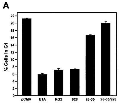
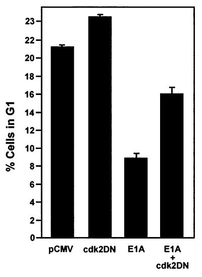
E1A requires p21 and the restoration of Cdk2 activity to overcome G1 arrest induced in p21+/+ cells after doxorubicin treatment. (A) p21+/+ cells were transfected in parallel with pCMV-E1A12S, E1A.RG2, E1A.928, E1A.dl26-35, E1A.dl26-35/928, or the control plasmid pCMV. Doxorubicin was added to the cultures immediately after, and at 36 h posttransfection, the cells were collected and stained with propidium iodide and anti-E1A for E1A expression. The cell cycle distribution of these cells was assessed by flow cytometry, as described in the legend to Fig. 4. (B) p21+/+ cells were transfected or cotransfected in parallel with a control plasmid (pCMV) and with pCMV and pCMV-E1A12S (4:1) or with Cdk2DN and pCMV-E1A12S (4:1), respectively. Afterwards, the cells were treated with doxorubicin, and at 36 h posttransfection, the cells were collected and processed for flow cytometry as described for panel A. The inset shows the expression of E1A and the coexpression of E1A and Cdk2DN in the doxorubicin-treated cells, as judged by Western blot analysis and the use of anti-E1A and anti-Cdk2. The histogram under the flow cytometry plots shows the percentage (+ standard deviation) of cells in a G1 population and is representative of three independent experiments.


