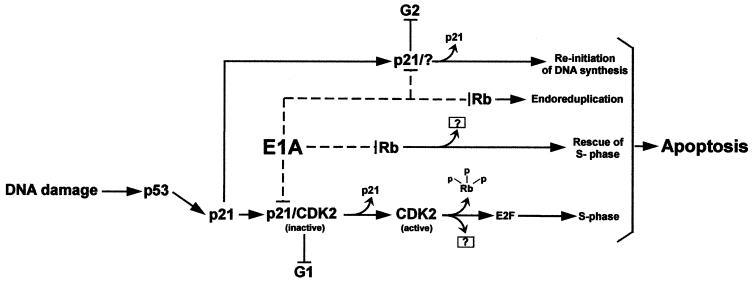
FIG. 8.
Model of E1A-induced apoptosis in DNA-damaged HCT116 cells. Exposure to doxorubicin results in the activation of p53 and, consequently, an increase in the levels of p21. In this cell system, the induced inhibitor (p21) antagonizes Cdk2 activity, leading to G1 arrest, and presumably functions in maintaining G2 arrest as well. Once E1A is introduced into the arrested cells, it targets p21, resulting in the restoration of Cdk2 activity. This in turn completes the phosphorylation of Rb and any other downstream targets that may be involved in promoting inappropriate entry into S phase, followed by apoptosis. The release of E2F from Rb has not been demonstrated in this system, and the model only considers its separate function in inducing cell cycle progression and not apoptosis. The model also suggests a likely but distinct role for E1A in inducing apoptosis by binding directly to Rb. The release of G2 and subsequent apoptosis as a function of E1A's direct effect on p21 in this pathway is speculative, as are the other components of the model which are indicated with question marks.

