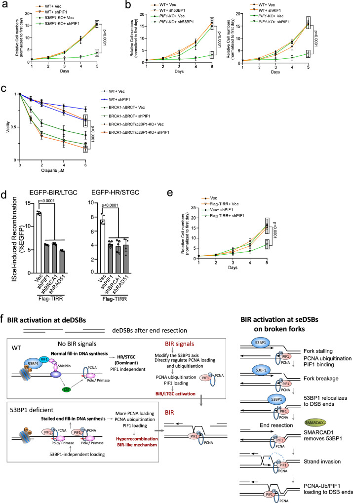
Fig. 7. Inactivation of BIR by targeting PIF1 causes cell death when the 53BP1 pathway is compromised.
a The growth curves of U2OS WT and 53BP1-KO cells were plotted after infection with lentiviruses expressing PIF1 shRNA with vector as a control. The expression of PIF1 was examined by qPCR (Supplementary Fig. 10a). (n = 3 replicates). b The growth curves of U2OS WT and PIF1-KO cells were plotted after infection with lentiviruses expressing shRNAs targeting 53BP1 (left) or RIF1 (right) with vector as a control. The expression of 53BP1 and RIF1 was examined by qPCR (Supplementary Fig. 10c). (n = 3 replicates). c Cell viability was determined in U2OS WT, BRCA1-ΔBRCT and 53BP1-KO/BRCA1-ΔBRCT cells expressing PIF1 shRNA with a vector control after treatment with the indicated concentrations of Olaparib for 72 hours. The expression of PIF1 was determined by qPCR (Supplementary Fig. 11a). (n = 3 replicates). d U2OS (EGFP-BIR/LTGC) cells (left) and U2OS (EGFP-HR/STGC) cells (right) overexpressing Flag-TIRR were infected with shRNAs targeting PIF1, BRCA1 or RAD51 with vector as a control, followed by infection with lentiviruses encoding I-SceI. The percentage of EGFP-positive cells was determined by FACS, 5 days post-infection. The expression of Flag-TIRR was examined by Western blot analysis (Supplementary Fig. 12a). The expression of PIF1, BRCA1 and RAD51 was determined by qPCR (Supplementary Fig. 12b). (n = 5 replicates). e The growth curves of U2OS cells with or without overexpressing Flag-TIRR were plotted after infection with lentiviruses expressing PIF1 shRNA with vector as a control. The expression of PIF1 was determined by qPCR (Supplementary Fig. 12c). (n = 3 replicates). Source data are provided as a Source data file. f Working models depicting the involvement of 53BP1 in limiting BIR at deDSBs (left) and at seDSBs on broken forks (right). See details in the main context.

