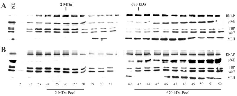
FIG. 2.
Gel filtration chromatography of RNAP II and GTFs. Nuclear extracts from uninfected cells (A) or cells infected with wild-type HSV-1 (B) were chromatographed on Sepharose CL-2B columns. Fractions were concentrated and analyzed by immunoblotting. Blots were probed with antibodies that recognize the RNAP II large subunit, the p56E subunit of TFIIE, the TBP subunit of TFIID, and the cdk7 subunit of TFIIH. Blots were also probed with an antibody that recognizes the DNA repair protein MLH1. Lane 1 contains 20 μg of uninfected (A) or infected (B) nuclear extract. Fractions 21 to 31 surrounded ∼2 MDa; fractions 42 to 52 were at and below ∼670 kDa. Ten fractions between approximately 1 MDa and 670 kDa are not shown in this figure. Fractions 24 through 28 (2 MDa Pool) were pooled for GST-TFIIS affinity chromatography and in vitro transcription assays (Fig. 4, 6, and 10A), as were fractions 43 through 52 (670 kDa Pool) (Fig. 7 and 10B).

