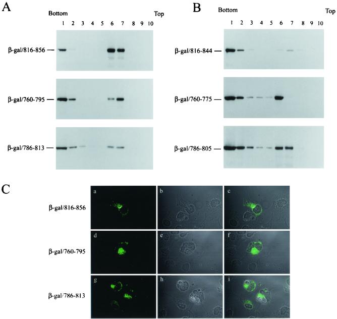
FIG. 4.
Each of the three LLP motifs confers membrane-targeting ability. (A) The membrane-targeting ability of the LLP sequences determined by membrane flotation assay. pCDNA3-β-gal chimeras each encoding sequences encompassing the LLP-1, LLP-2, or LLP-3 motif as indicated were assessed by membrane flotation assay. (B) Effects of deletions in the C terminus of the LLP motifs on membrane binding. Postnuclear supernatants obtained from COS-1 cells expressing sequences containing deletions in the C terminus of LLP-1, LLP-2, and LLP-3, respectively, were analyzed by sucrose gradient equilibrium centrifugation. (C) Subcellular localization of the β-galactosidase/LLP fusion proteins. COS-1 cells expressing β-galactosidase/LLP fusion proteins were immunostained with β-galactosidase MAb and FITC-conjugated anti-mouse immunoglobulin G and then were examined by confocal microscopy as shown in panels a, d, and g, respectively. The phase-contrast images for the fields examined are shown in panels b, e, and h, respectively. The immunofluorescence staining and phase-contrast images were superimposed, and the images are shown in panels c, f, and i, respectively.

