Fig. 1.
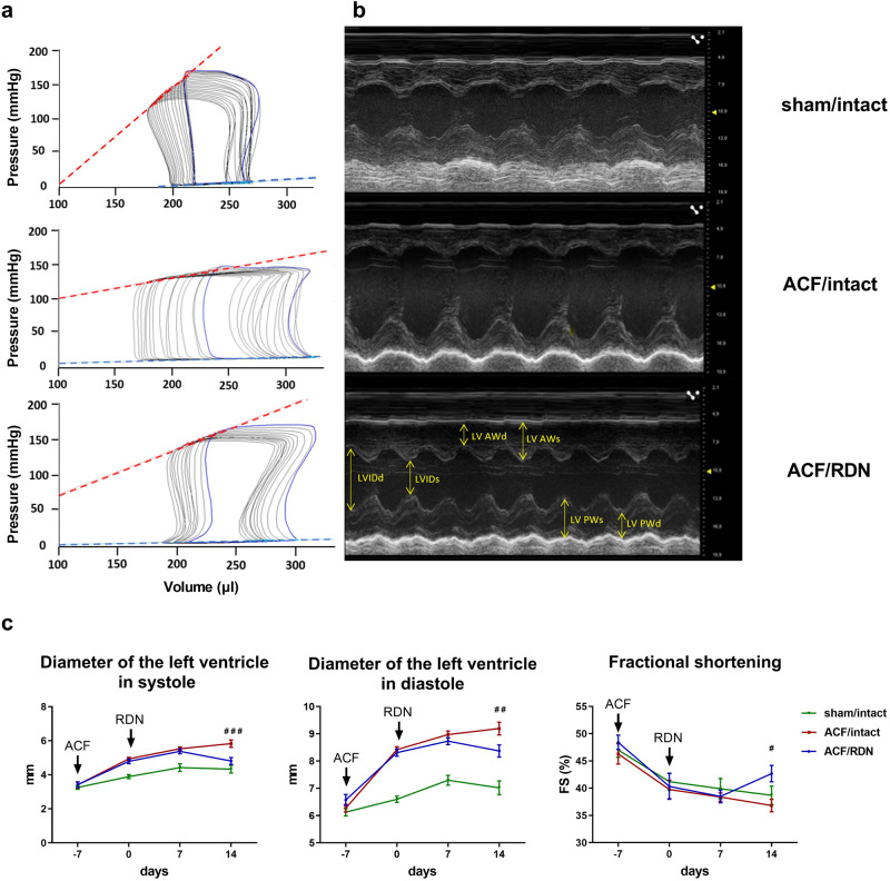
In vivo measurement of LV contractility and dimensions. a Representative pressure-volume loops from invasive pressure-volume analysis. Red line—end-systolic elastance (Ees), blue line—end-diastolic pressure-volume relationship (EDPVR). b Echocardiographic M mode images of parasternal long axis view. LV AWd left ventricular anterior wall thickness in diastole, LV AWs left ventricular anterior wall thickness in systole, LVIDd left ventricular internal diameter in diastole, LVIDs left ventricular internal diameter in systole, LV PWd left ventricular posterior wall thickness in diastole, LV PWs left ventricular posterior wall thickness in systole. c Diameter of left ventricle in systole (LVIDs) and diastole (LVIDd) measured during each week of experiment (3 weeks); FS fractional shortening. N = 10 in sham/intact, N = 19 in ACF/intact, N = 13 in ACF/RDN. ###p < 0.001; ##p < 0.01; #p < 0.05, ACF/intact vs. ACF/RDN group, compared to the day 14

