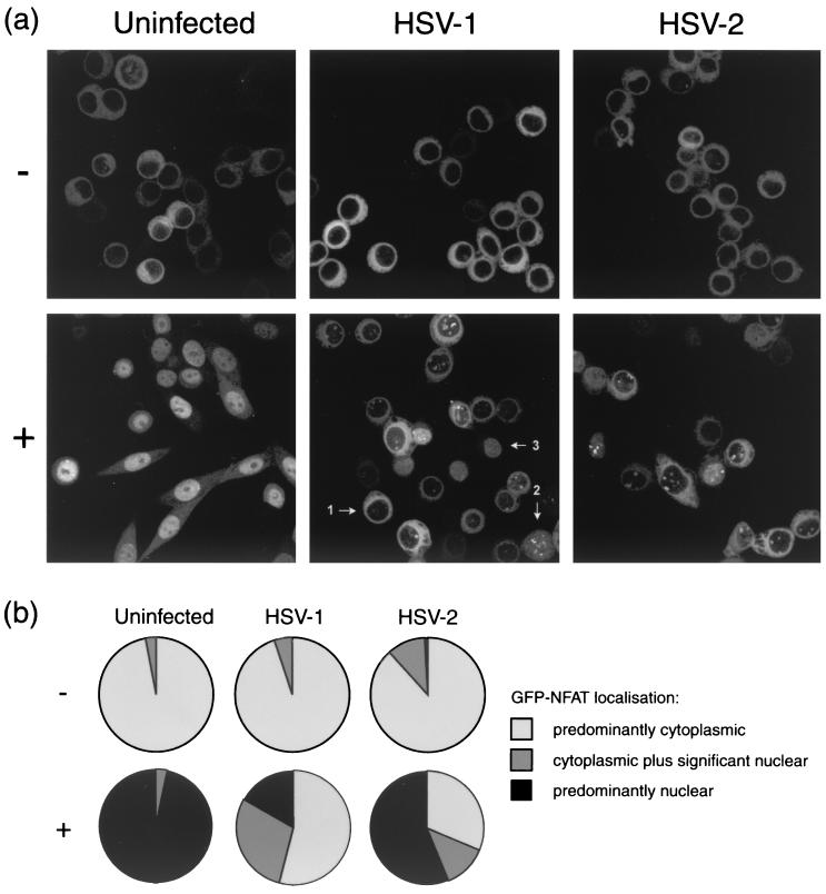
FIG. 1.
HeLa cells stably expressing GFP-NFAT were infected with either HSV-1 or HSV-2 or left uninfected. At 5 h, cells were treated with 1 μM ionomycin–30 mM lithium acetate (+) or left untreated (−). At 7 h, cells were fixed in methanol and examined by confocal microscopy. (a) Representative images of each experimental condition as described in the text. (b) Summary of GFP-NFAT localization, scored for every cell in five randomly chosen fields of view in each experimental condition. Cells were scored as having GFP-NFAT localization that was predominantly cytoplasmic, cytoplasmic plus significantly nuclear, or predominantly nuclear, and examples of each cell type (arrows 1, 2, and 3, respectively) are marked in the panel for treated, HSV-1-infected cells in panel a. Approximately 100 cells were counted for each of the test conditions.

