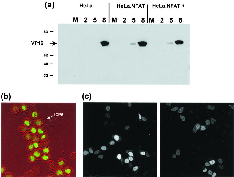
FIG. 2.
HeLa cells or HeLa cells stably expressing GFP-NFAT were mock infected (M) or infected with HSV-1 without or with (+) pretreatment with trichostatin. (a) At 2, 5, or 8 h, as indicated, infected cells were harvested and the accumulation of VP16 was measured by SDS-polyacrylamide gel electrophoresis followed by Western blotting with an anti-VP16 monoclonal antibody. Numbers on the left are molecular masses in kilodaltons. (b) HeLa cells stably expressing GFP-NFAT infected as for panel a in the presence of trichostatin were fixed 12 h after infection, and the accumulation of the late viral protein ICP5 was analyzed by immunofluorescence. The localization of ICP5 (green channel) is superimposed on the phase image of the infected cells to emphasize ICP5 nuclear accumulation. (c) HeLa cells transiently expressing GFP.HCF.NLS were infected with HSV-1 (right panel) or left uninfected (left panel), and localization in live cells was examined by confocal microscopy at 5 h postinfection.

