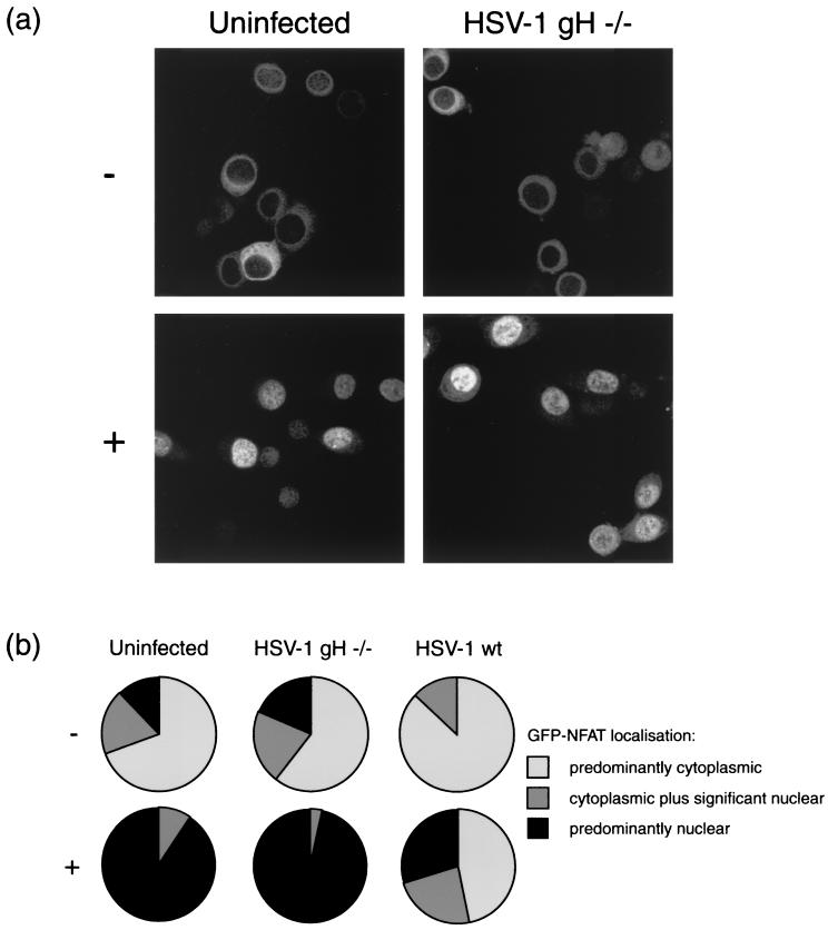
FIG. 3.
HeLa cells stably expressing GFP-NFAT were infected with either HSV-1 or ΔgH HSV-1 or left uninfected. At 5 h, cells were treated with 1 μM ionomycin–30 mM lithium acetate (+) or left untreated (−). At 7 h, cells were fixed in methanol and examined by confocal microscopy. (a) Representative images of treated or untreated uninfected and ΔgH HSV-1-infected cells. (b) Summary of GFP-NFAT localization, scored for every cell in five randomly chosen fields of view in each experimental condition.

