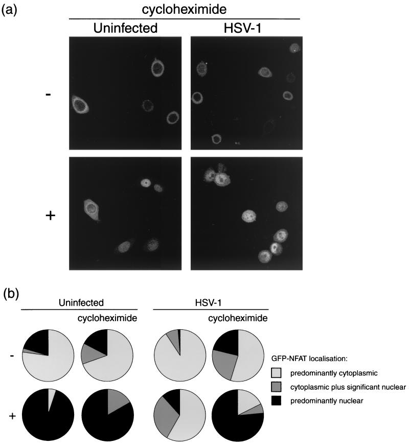
FIG. 4.
HeLa cells stably expressing GFP-NFAT were infected with HSV-1 or left uninfected. At 5 h, cells were treated with 1 μM ionomycin–30 mM lithium acetate (+) or left untreated (−). Each assay was performed in duplicate, with one set of cells being examined in the presence of 50 μg of cycloheximide per ml, added at 30 min prior to infection. At 7 h, cells were fixed in methanol and examined by confocal microscopy. (a) Representative images of treated or untreated uninfected and infected cells in the presence of cycloheximide. (b) Summary of GFP-NFAT localization, scored for every cell in five randomly chosen fields of view in each experimental condition.

