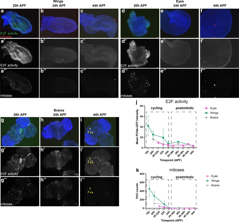
Fig. 1.
The timing of cell cycle exit in the Drosophila wing, eye and brain are similar. (a–c) Wings, (d–f) eyes, and (g–i) brains were dissected from staged pupa and stained for mitoses (anti-Phospho-Histone H3) and E2F transcriptional activity (anti-GFP for PCNA-GFP reporter) at the timepoints indicated. Quantifications of PCNA-GFP reporter and mitotic counts are provided in (j) and (k) respectively. Animals were collected as white pre-pupa (0 h APF) and incubated at 25°C to the indicated timepoints. Yellow arrowheads indicate 4 of the 8 mushroom body neuroblasts that continue to cycle until 96 h APF (Siegrist et al. 2010). In overlays, blue = DNA (Dapi), red = mitoses (PH3), and green = E2F activity (PCNA-GFP reporter). Sample numbers; eyes 20–22 h n = 5, 24 h n = 5, 36–44 h n = 7, wings 18–22 h n = 12, 24 h n = 2, 36–44 h n = 2, brains, 20–22 h n = 14, 24 h n = 7, 36–44 h n = 5.

