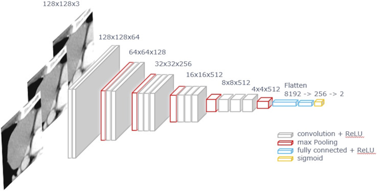
Figure 2.
Convolutional neural network model. The center slice and pre- and post-slices from CT images of thymomas in the axial direction were used as three channel inputs, and training was performed using a 2-dimensional convolutional neural network, which was a fine-tuning model based on VGG16. CT, computed tomography.

