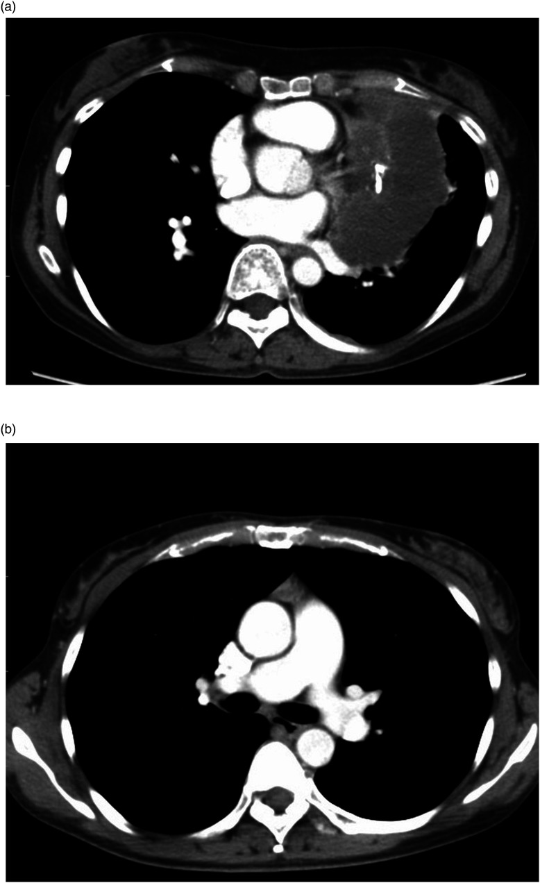
Figure 3.
(a) A 47-year-old woman with a low-risk thymoma (type B1). The DL model correctly classified it as a low-risk thymoma, although all three radiologists classified it as high-risk thymoma. (b) A 46-year-old woman with a high-risk thymoma (type B2). The DL model correctly classified it as a high-risk thymoma, although all three radiologists classified it as low-risk thymoma.

