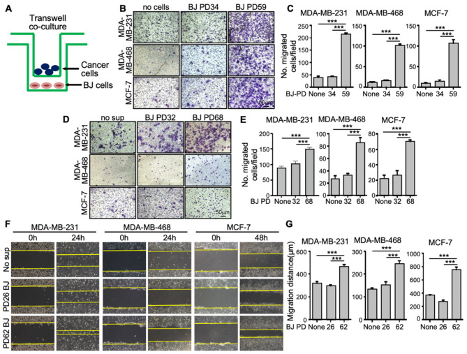
Fig. 1.
Senescent BJ cells promote the motility of breast cancer cells
(A) Schematic diagram of the co-culture system in which young or senescent BJ cells and breast cancer cells were seeded in the lower and upper chambers, respectively, of the transwells. The 2 chambers were separated by a membrane with 8-micron pores. Migration of the breast cancer cells towards the lower chamber was measured. (B-C) Representative crystal violet-stained images of transwell migration of MDA-MB-231, MDA-MB-468 and MCF-7 cells co-cultured with medium (no cells or None) or young (PD34) or senescent (PD59) BJ cells for 12 h (B), and quantification of number of migrated cells per 20X field (mean ± SD, n = 3) (C). At least 5 randomly chosen 20X fields were counted for each of the triplicates. (D-E) Representative crystal violet-stained images of transwell migration of MDA-MB-231, MDA-MB-468 and MCF-7 cells co-cultured with medium only (no sup or None) or conditioned medium from young (PD32) or senescent (PD68) BJ cells for 16 h (D), and quantification of number of migrated cells per 20X field (mean ± SD, n = 3) (E). At least 5 randomly chosen 20X fields were counted for each of the triplicates. (F-G) Representative images of MDA-MB-231, MDA-MB-468 and MCF-7 cells immediately (0 h), 24 h (24 h, for MDA-MD-231 and MDA-MB-468) or 48 h (48 h, for MCF-7) after a scratch wound was made and co-cultured with medium only (no sup or None) or conditioned medium from young (PD26) or senescent (PD62) BJ cells (F), and quantification of the distance (µM) of the wound edges by ImageJ at 24 h (for MDA-MD-231 and MDA-MB-468) or 48 h (for MCF-7) (mean ± SD, n = 3) (G). (C, E, G) *** p < 0.001 between indicated groups in One-way ANOVA corrected for multiple comparisons using Tukey’s multiple comparison correction adjustment

