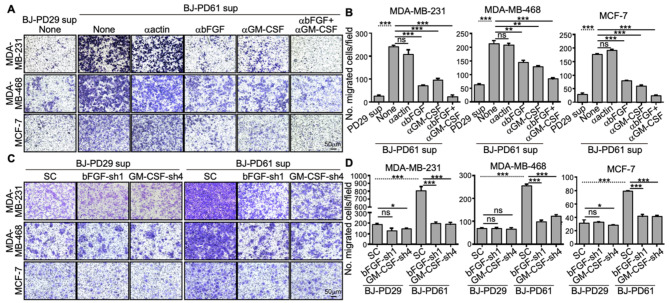
Fig. 3.
Senescent cells promote the migration of breast cancer cells via secreted bFGF and GM-CSF. (A-B) Representative crystal violet-stained images of transwell migration of MDA-MB-231, MDA-MB-468 and MCF-7 cells co-cultured with conditioned medium from young (PD29) or senescent (PD61) BJ cells, which were left untreated (None) or incubated with neutralizing antibodies against β-actin (1 ng/ml), bFGF (1 ng/ml), GM-CSF (5 ng/ml) or both bFGF and GM-CSF for 12 h (A), and quantification of number of migrated cells per field (mean ± SD, n = 3) (B). At least 5 randomly chosen 20X fields were counted for each of the triplicates (C-D) Representative crystal violet-stained images of transwell migration of MDA-MB-231, MDA-MB-468 and MCF-7 cells co-cultured with conditioned medium from young (PD29) or senescent (PD61) BJ cells transduced with shRNA control (SC) or shRNAs for bFGF or GM-CSF for 20 h (C), and quantification of number of migrated cells per field (mean ± SD, n = 3) (D). At least 5 randomly chosen 20X fields were counted for each of the triplicates. (B, D) ns, not significant; * p < 0.05; ** p < 0.01; and *** p < 0.001 between indicated groups in unpaired, 2-sample t tests (dotted lines) or One-way ANOVA corrected for multiple comparisons using Tukey’s multiple comparison correction adjustment (solid lines)

