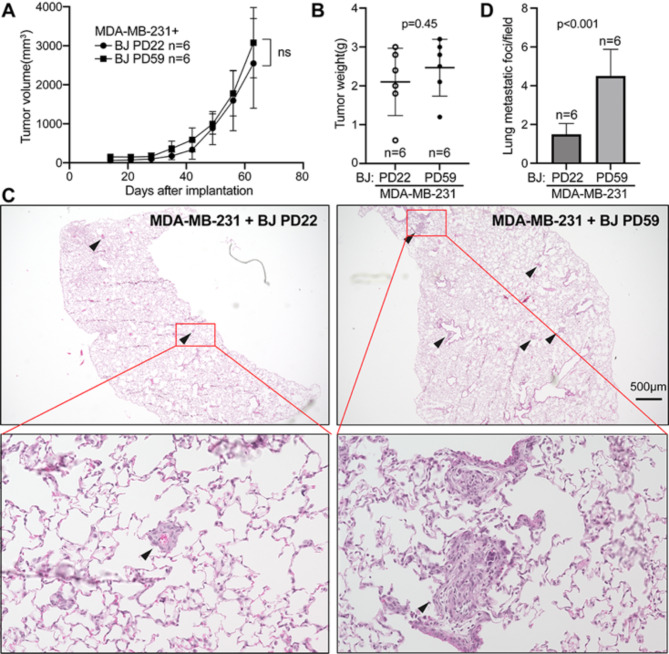
Fig. 6.
Senescent cells promote breast cancer metastasis in xenograft tumor models. 5 × 10⁵ of MDA-MB-231 cells and 5 × 10⁵ of young (PD22) or senescent (PD59) BJ cells were co-injected into the mammary fat pads of 6–8-week-old nude mice. Tumor sizes were measured weekly over 9 weeks. Tumor growth curves were plotted (A). Upon sacrifice, tumors were removed and weighted (B), and lung sections were stained by hematoxylin and eosin, photographed (C) and quantified for the number of metastatic foci (indicated by arrows) (D). In (C), images of the representative lung sections with metastatic foci (indicated by arrows) are shown in the top panels, and one metastatic focus (indicated by red boxes) from each section is magnified and shown in the bottom panels. In (D), for each mouse among the total of 6 in each group, the number of metastatic foci were counted in 5 randomly chosen 2X fields in a section containing all 5 lung lubes, and the number of metastatic foci per field was calculated. (A-B, D) Values are mean ± SD, n = 6 mice per group. p values are from unpaired, 2-sample t tests. ns, not significant

