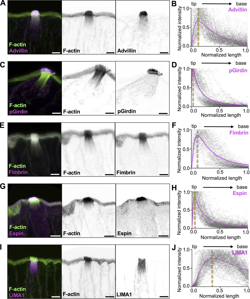
Figure 4.
Tuft cell actin-binding proteins exhibit regionalized localization along the core bundle axis. (A, C, E, G, and I) MaxIP Airyscan image of lateral frozen tissue section and immunostained for actin-binding proteins, (A) advillin, (B) pGirdin, (C) fimbrin, (D) Espin, and (E) LIMA1 in addition to actin marked with phalloidin (scalebar = 5 µm). (B, D, F, H, and J) Graph of linescans drawn from MaxIP images from apical tip to the base of core bundle, measuring the intensity of actin-binding proteins. Raw values depicted in gray, line fit (lognormal) in magenta; the yellow line indicates the peak of line fits (n = advillin, 29 tuft cells; pGirdin, 32 tuft cells; fimbrin, 30 tuft cells; espin, 33 tuft cells; LIMA1, 32 tuft cells, over 3 mice).

