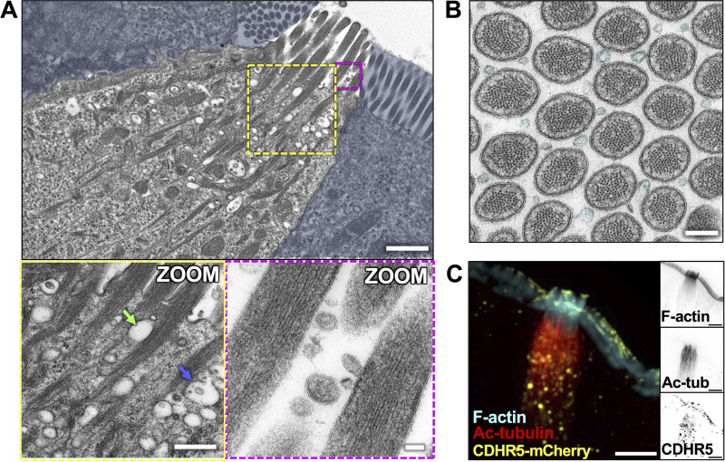
Figure 7.
Membranous organelles are associated with the tuft cell cytoskeletal network. (A) TEM of ultrathin tissue slice showing lateral tuft cell section (scalebar = 1 µm) with enterocytes masked in blue. Zoom inset (left) shows vesicles along core bundles near the apical surface (scalebar = 400 nm). Blue arrow points to an MVB, green arrow points to an electron-lucent vesicle. Zoom inset (right) shows EVs between apical protrusions (scalebar = 50 nm). (B) TEM of ultrathin tissue slice showing en face section of the apical tuft showing EVs (cyan) between protrusions (scalebar = 200 nm). This image was taken of the same tuft cell shown in Fig. 2 A. (C) MaxIP SDC image of tuft cell from CDHR5-mCherry mouse, immunostained for mCherry to boost the signal and acetylated tubulin (scalebar = 5 µm).

