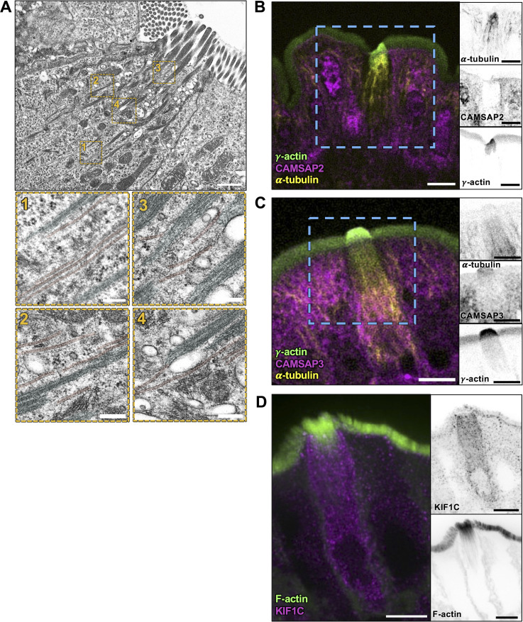
Figure S5.
Microtubules align with core actin bundles but lack clear polarity markers in tuft cells. (A) TEM of ultrathin tissue slice showing lateral tuft cell section (scalebar = 1 µm) also shown in Fig. 7 A. Below, four zoomed areas show microtubules, pseudo-colored red, in proximity of the core actin bundle rootlets, pseudo-colored cyan (scalebar = 200 nm). (B and C) MaxIP SDC image of paraffin-embedded tissue and immunostained for (B) CAMSAP2 and (C) CAMSAP3 with microtubules marked by staining for α-tubulin and actin marked with staining for γ-actin (scalebar = 5 µm). (D) MaxIP SDC image of frozen tissue section immunostained for KIF1C with F-actin marked by phalloidin staining.

