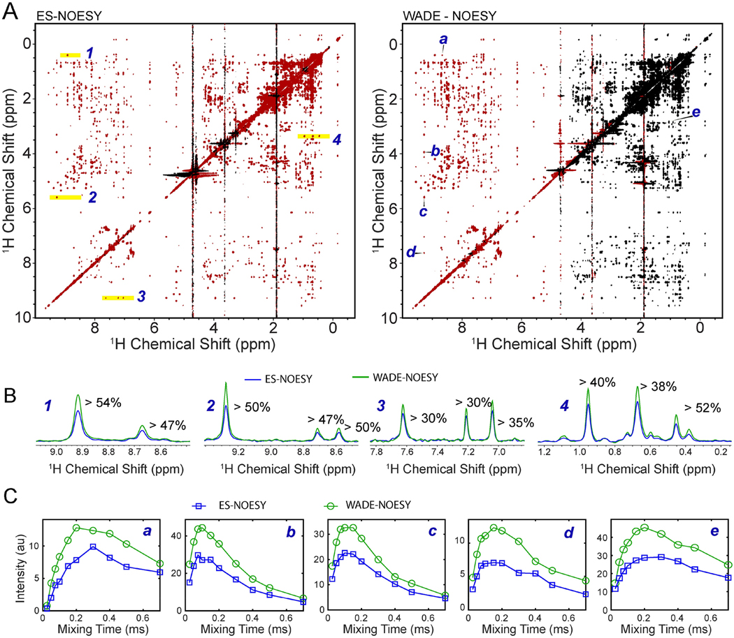
Figure 6.
(A) Comparison of the 2D [1H-1H] NOESY of UBIK48C with a 150 ms mixing time using ES and WADE suppression scheme. The peaks in red are positive and those in black are negative. All experiments were performed on a Bruker 850 MHz spectrometer at 303 K using identical acquisition parameters: 256 number of complex points were acquired in indirect dimension and 8 scans per FID with a relaxation delay of 2 sec. Identical processing parameters were used for both spectra. (B) 1D slices of the 2D [1H-1H] ESNOESY (blue) and [1H-1H] WADE-NOESY (green) for the peaks highlighted in A. (C) NOESY buildup curves [1H-1H] ES-NOESY(blue) and [1H-1H] WADE-NOESY (green) for the resonances marked A.

