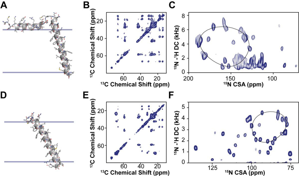
Figure 1: MAS and OS-ssNMR studies of single pass membrane proteins PLN and SLN.
A. Structure and membrane orientation of PLN obtained from a combination of isotropic and anisotropic restraints. B. 13C,13C-DARR spectrum of PLN in lipid vesicles. C. SE-SAMPI4 spectrum of PLN in oriented lipid bicelles. D. Structure and orientation of SLN in lipid membranes calculated using both MAS and OS-ssNMR data. E. 13C,13C-DARR spectrum of SLN in lipid vesicles. F. SE-SAMPI4 spectrum of PLN in oriented lipid bicelles. Note that the oriented spectrum of SLN was obtained using paramagnetic doping and in the absence of Yb3+ (unflipped bicelles).

