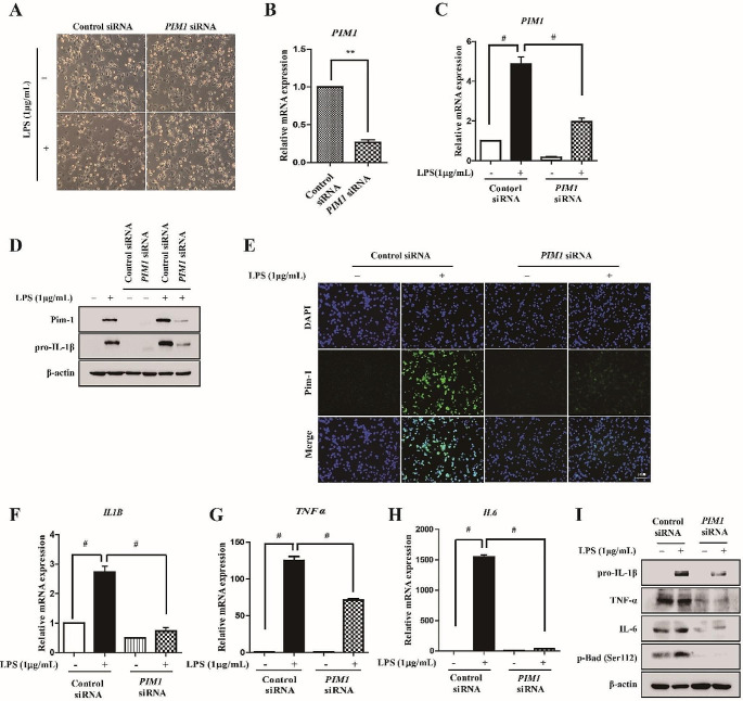
Fig. 2.
The effect of PIM1 knockdown in LPS-mediated inflammatory signals of macrophage-like THP-1 cells. THP-1 cells transfected with control siRNA or PIM1 siRNA for 72 h and then stimulated with LPS (1 µg/mL) for 6 h. (A) The effects of PIM1 siRNA on cellular morphological changes were observed by microscopy (×200). (B, C) Total RNA was extracted, and used to evaluate the mRNA expression levels of PIM1 by real-time qPCR, (** p < 0.01, # p < 0.001). (D) Whole cell lysates were isolated and used to measure the protein expression levels of Pim-1 and pro-IL-1β by Western blotting. (E) Cells were stained with antibodies to Pim-1 (green) and DAPI (blue) and captured at ×200 using fluorescence microscope (scale bar = 50 μm). (F-H) Total RNA was extracted, and used to evaluate the mRNA expression levels of pro-inflammatory cytokines (IL1B, TNFα, and IL6) (# p < 0.001). (I) Whole cell lysates were isolated and used to measure the protein expression levels of pro-IL-1β, IL-6, TNF-α, and p-Bad (Ser112) by Western blotting

