
Figure 3. Cannabinoid-LTD occurs at AIC-DLS synapses.
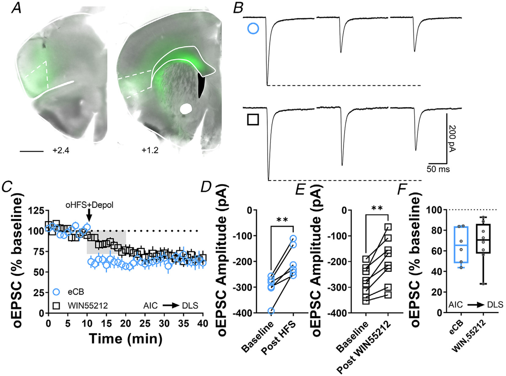
A, coronal brain slice showing the AAV-ChR2 infection of AIC and dorsal striatal terminal expression (bar scale = 1000 μm). B, representative oEPSC traces showing the effects of pairing optical high-frequency stimulation with postsynaptic depolarization (oHFS+Depol, cyan open circle) to physiologically induce LTD, presumably mediated by endocannabinoids and the application of the CB1 agonist, WIN55,212-2 (1 μM, 10 min, black open square) to chemically induce CB1-LTD in brain slices of AAV-ChR2 C57Bl/6J mice. C, both oHFS and WIN55,212-2 induced AIC-DLS LTD. Data represent means ± SEM. D, oHFS significantly reduced oEPSC amplitudes (0–10 min baseline vs. final 10 min of recording; paired t test, P = 0.00350, t5 = 5.2, n = 6 neurons from four mice). E, WIN55,212-2 significantly reduced oEPSC amplitudes (0–10 min baseline vs. final 10 min of recording; paired t test, P = 0.00179, t7 = 4.9, n = 8 neurons from four mice). F, the box plot shows the final 10 min of recording average of stimulation-induced LTD (65 ± 7%) and CB1-LTD (68 ± 7%). **P < 0.01.