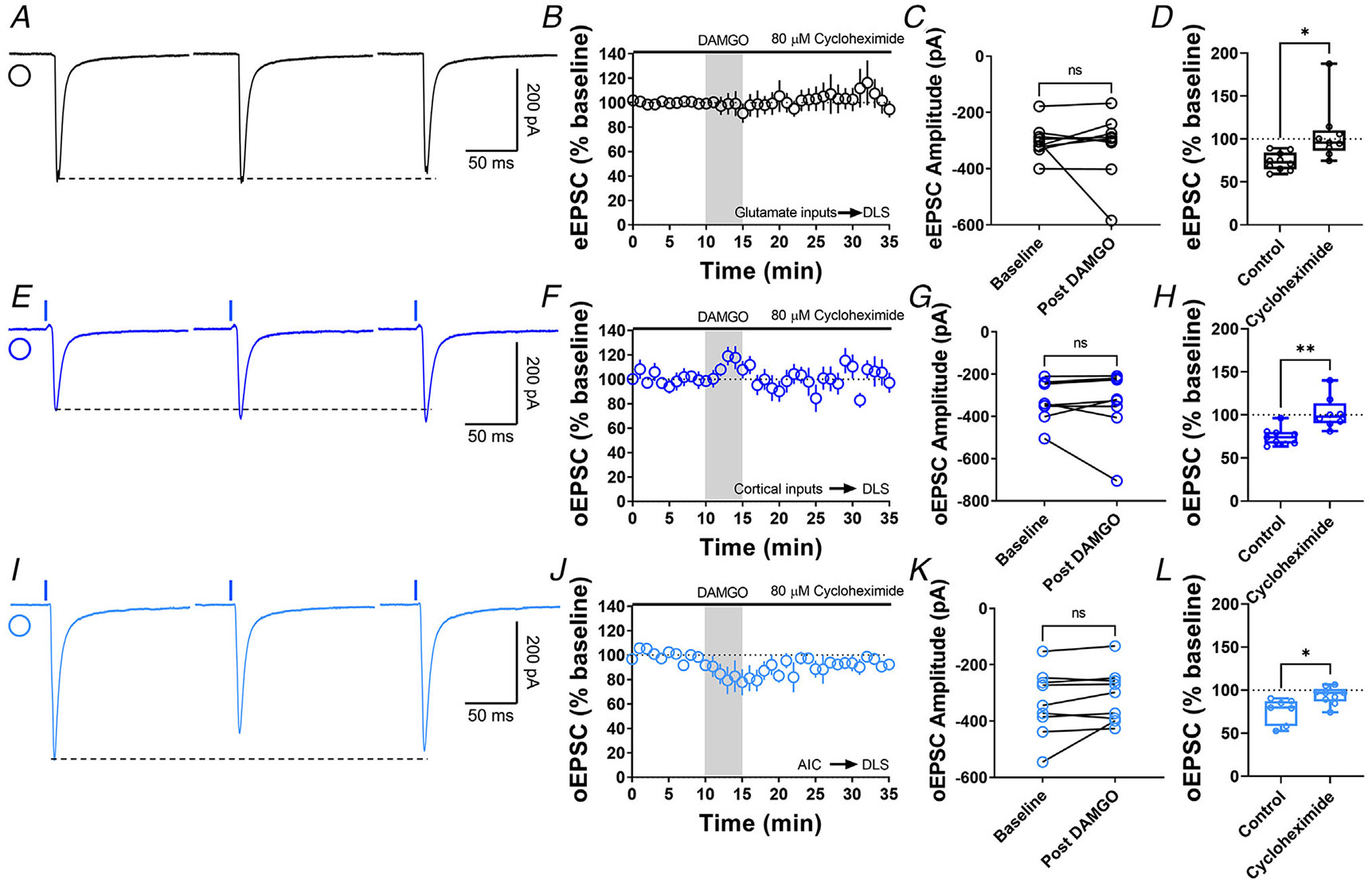
Figure 7. Protein translation is required for MOR-mediated LTD expression.

A, representative eEPSC traces showing the effects of 80 μM cycloheximide before, during and after DAMGO (0.3 μM, 5 min) application. B–D, protein translation inhibition disrupted glutamatergic MOR-LTD (105 ± 11%, unpaired Welch’s t test, P = 0.0228, t10 = 2.72), showing no changes in eEPSC amplitude after DAMGO bath application (0–10 min baseline vs. final 10 min of recording; paired t test, P = 0.651, t8 = 0.471, n = 9 neurons from four mice). E, representative oEPSC traces showing the effects of 80 μM cycloheximide before, during and after DAMGO (0.3 μM, 5 min) application in brain slices from Emx1-Ai32 mice. F–H, protein translation inhibition blocked corticostriatal MOR-LTD (102 ± 7%, unpaired t test, P = 0.00163, t15 = 3.83) with no effects in oEPSC amplitude after DAMGO bath application (0–10 min baseline vs. final 10 min of recording; paired t test, P = 0.633, t7 = 0.499, n = 8 neurons from three mice). I, representative AIC-DLS oEPSC traces showing the effects of 80 μM cycloheximide before, during and after DAMGO (0.3 μM, 5 min) application. J–L, protein translation is needed to induce MOR-LTD (94 ± 3%, unpaired t test, P = 0.0129, t14 = 2.85) from AIC inputs. DAMGO did not change in oEPSC amplitude (0–10 min baseline vs. final 10 min of recording; paired t test, P = 0.163, t8 = 1.54, n = 9 neurons from three mice). Time course data represent means ± SEM. Box plots show average of the final 10 min of recording and represent median and interquartile ranges. ns = not significant, *P < 0.05, **P < 0.01.
