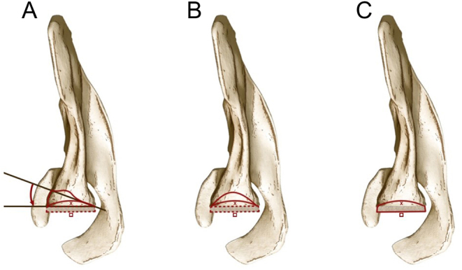
Figure 3.

Drawing of the three situations, as viewed in the axial plane, in which glenoid bone grafting was performed in the included studies. (A) Glenoid version correction (curved red arrow) and lateralization (red square) of the COR from its native position (red x mark) in situations of uncontained glenoid bone defects; (B) Glenoid bone grafting (continuous red line) and lateralization of the COR (red square) from its native position (red x mark) in cases of contained glenoid defects; (C) Glenoid COR lateralization (red square) from its native (red x mark) in patients with minimal glenoid bone erosion.

 This work is licensed under a