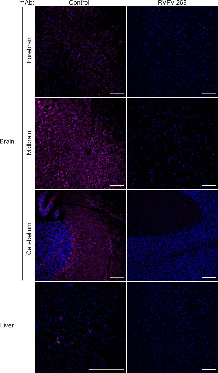
Figure 3. RVFV is less diffuse or not observed in the brains of rats pretreated with mAb RVFV-268.
Immunofluorescence images of the forebrain, midbrain, cerebellum, and liver stained for nuclei (DAPI; blue) and RVFV nucleoprotein (magenta) from the brain or liver of rats pretreated with control antibody (left) or RVFV-268 (right). Original magnification, ×20. Scale bar: 100 μm (first 3 rows and bottom right); 200 μm (bottom left).

