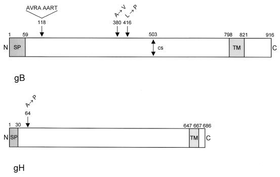
FIG. 8.
Diagram of mutations present in gB Pass and gH Pass. (Top) Schematic representation of wild-type gB (38). Mutations in gB Pass are indicated above. (Bottom) Schematic representation of wild-type gH (18). The sole mutation in gH Pass is indicated above. N, amino terminus; SP, signal peptide; TM, transmembrane domain; C, carboxy terminus; CS, furin protease cleavage site in gB.

