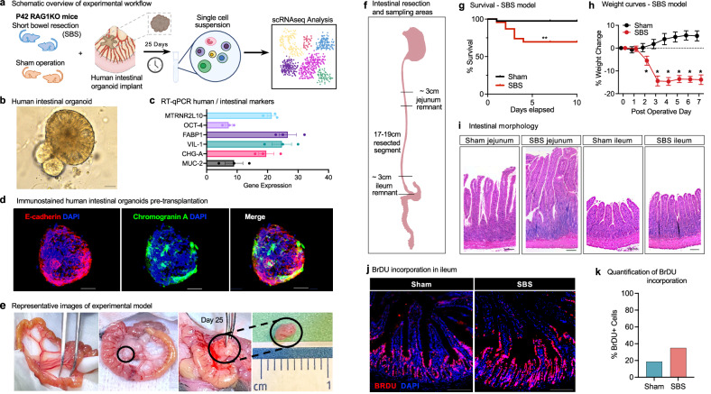
Fig. 1. Development of a human–mouse chimera model system for the identification of pathways that mediate intestinal adaptation in experimental SBS.
a Schematic overview of the experimental workflow for the human small intestinal organoid xenotransplantation SBS model. Six-week-old Rag1 KO C57BL/6 mice underwent experimental SBS (n = 5) or sham surgery (n = 5). Human intestinal organoids, differentiated from iPSCs, were implanted into the mesentery using surgical glue. Xenotransplants were harvested after 25 days for single-cell RNA sequencing analysis. b Bright field microscopy of human small intestinal organoids differentiated from iPSC (Day 42). Magnification ×20. Scale bar 100 μm. c RT-qPCR analysis of intestinal-specific markers in human organoids pre-xenotransplantation, showing increased expression relative to housekeeping gene RPLP0. Data from three independent organoid cultures are presented as mean ± standard error of the mean (SEM). d Immunofluorescence staining of pre-xenotransplantation organoids for epithelial and intestinal markers. Scale bar 100 µm. e Images of mouse intestine pre-resection, during xenotransplantation, and at harvest on Day 25. f Schematic of intestinal resection and sampling areas in the SBS model. 75% of the small bowel was resected, with a jejuno-ileal anastomosis. Sham controls underwent transection and anastomosis without resection. g Survival curves for SBS (n = 23) and sham (n = 40) mice with human xenotransplants. SBS mice had ~70% survival at 10 days vs ~100% for sham (log-rank test, p = 0.0015). h Growth curves showing significant weight loss in SBS mice (n = 6) compared to steady weight gain in sham controls (n = 7). Weight differences were significant at all indicated time points (t-test, *p < 0.05). Data are mean ± SEM. i H&E staining of jejunum and ileum from Rag1 KO mice post-xenotransplant. SBS jejunal villi were significantly lengthened; ileal villi were modestly increased. Scale bar 100 µm. j Immunofluorescence images of BrdU staining in intestinal tissue post-xenotransplant. BrdU incorporation was higher in SBS ileum compared to sham. Scale bar 100 µm. k Quantification of BrdU-positive cells in intestinal crypts showed significantly higher incorporation in SBS compared to sham controls. Source data are provided as a separate file.

