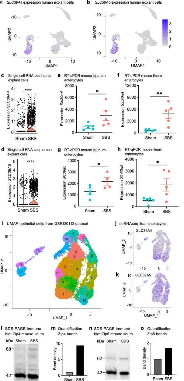
Fig. 3. Zinc transport genes SLC39A4 and SLC39A5 are upregulated in SBS.
UMAP visualization of human small intestinal xenotransplants from five SBS mice and five sham controls. Higher expression of SLC39A4 (a) and SLC39A5 (b) is observed within the enterocyte 1 cluster derived from SBS. Violin plots depict the elevated expression of SLC39A4 (c) and SLC39A5 (d) in human SBS xenotransplants compared to sham. Wilcoxon rank sum test, ****p < 0.0001. RT-qPCR of isolated intestinal epithelial cells of SBS mice (n = 5) shows the increased relative expression of Slc39a4 in the jejunum (e) and ileum (f) and of Slc39a5 expression in SBS jejunum (g) and ileum (h) compared to sham control (n = 5). Mann–Whitney U-test. *p ≤ 0.05; **p ≤ 0.01. Data represent mean ± SEM. i UMAP projection of processed, filtered, and clustered SBS epithelial cell data from the gene expression omnibus GSE130113 generated in Seurat. Feature plots illustrate the expression of j SLC39A4, and k SLC39A5 genes in mouse SBS native epithelial cells in GSE130113. Representative Western blot analysis of Zip4 (l) and Zip5 (m) protein expression in sham and SBS mice. Quantification analysis of Zip4 (n) Zip5 (o) protein band density from blots comparing ileum tissue from sham and SBS mice fed a control diet. Source data are provided as a Source Data file.

