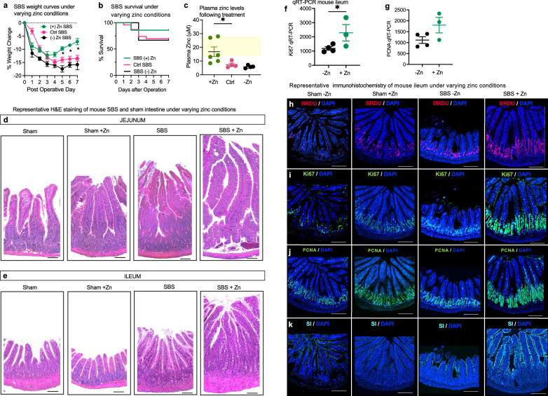
Fig. 5. Zinc supplementation in murine SBS enhances intestinal proliferation and adaptation in vivo.
a Growth curves for SBS mice treated with control (Ctrl SBS) (n = 10), zinc supplementation ((+) Zn SBS) (n-6), and zinc-depleted diet ((−) Zn SBS) (n = 6) over 7 days. Significant weight change is observed in (+) Zn SBS compared to Ctrl SBS from Day 5 onward. Data points are mean ± SEM. t-test, *p < 0.05 at indicated time points. b Survival curves comparing the three groups within the 7-day SBS model. c Plasma zinc levels measured by colorimetric assay (Abcam). Zinc-supplemented mice (n = 6) achieved plasma zinc levels within or above the normal range (12–25 µmol/l, yellow-shaded area). SBS mice on control (n = 4) and zinc-depleted diets (n = 4) did not reach this range. Data are mean ± SEM. t-test, p = 0.0336. H&E staining of the jejunum (d) and ileum (e) tissue following the 7-day SBS model. Mice were fed either a control or a zinc-supplemented diet. Magnification: 20x; scale bar: 100 µm. RT-qPCR analysis of ileal tissue from −Zn SBS mice (n = 4) vs +Zn SBS mice (n = 3), showing ~ 2-fold increase in Ki67 expression (f, p < 0.05) and a trend towards increased PCNA expression (g) in the zinc-supplemented group (p = 0.0540). Data are mean ± SEM. Representative immunofluorescence images of intestinal tissue from SBS mice following 10 days of post-operative treatment with zinc depleted or zinc-supplemented diet. h BRDU staining 24 h after gavage; Ki67 expression (i), PCNA expression (j), and SI expression (k) under the same conditions. Magnification 10×; scale bar 100 µm. Source data are provided as a Source Data file.

