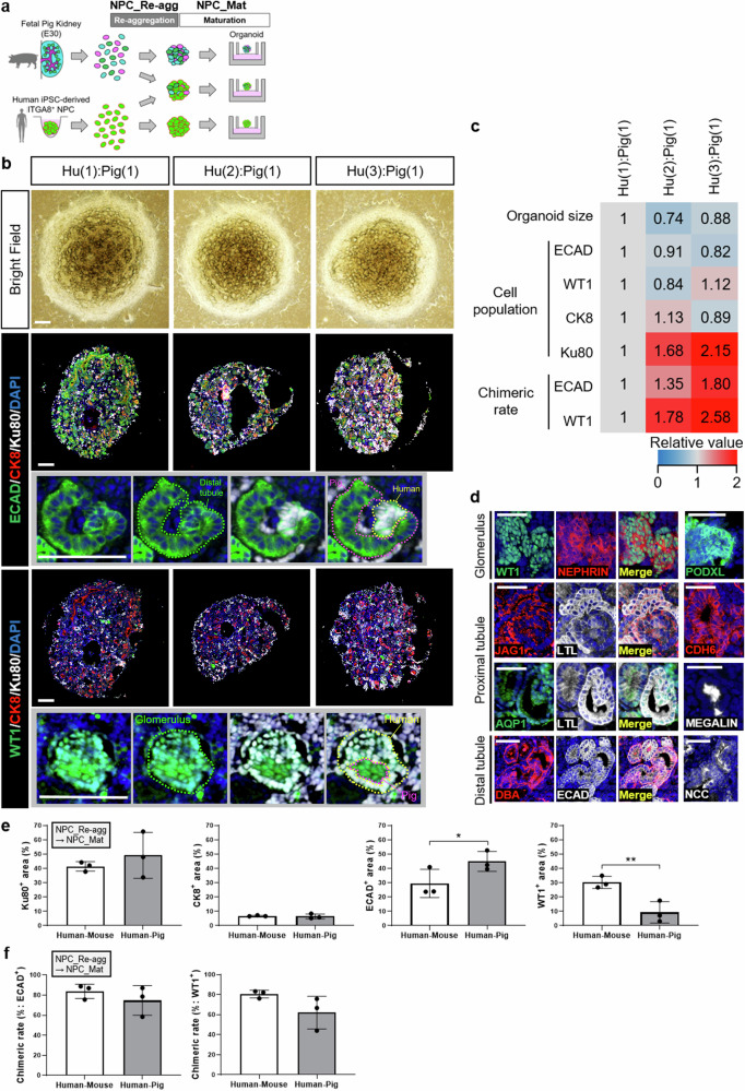
Fig. 4. Generation and evaluation of human-pig chimeric renal organoids.
a Experimental scheme for generating human-pig chimeric renal organoids using fetal pig kidneys and human iPSC-derived NPCs. b Bright-field images and immunostaining images of chimeric renal organoids formed at different human-pig mixing ratios and stained by a distal tubule marker, a glomerular marker, and a human cell marker. Scale bars represent 200 μm. c Quantitative analysis of size, cellular composition ratio, and chimeric structure formation rate using human-pig chimeric renal organoids at different human-pig cell mixing ratios. d Images of human(3)-pig(1) chimeric renal organoids immunostained by antibodies at various developmental stages of each nephron segment. Scale bars represent 100 μm. e Cellular composition analysis based on images of organoids after immunostaining. The quantitative data of human-pig chimeric renal organoids were obtained from chimeric organoid images with a human-pig cell mixing ratio of 3:1. The quantitative data of human-mouse chimeric renal organoids were reutilized from Fig. 2e (n = 3 independent experiments; mean ± s.d.; *P < 0.05, **P < 0.01; two-tailed t-test). f Quantitative analysis of chimera formation rate. The left graph indicates the chimera rate in images of stained distal tubules, while the right graph indicates the chimera rate in images of stained glomeruli. The quantitative data of human-pig chimeric renal organoids were obtained from chimeric organoid images with a human-pig cell mixing ratio of 3:1. The quantitative data of human-mouse chimeric renal organoids were reutilized from Fig. 2f (n = 3 independent experiments; mean ± s.d.; two-tailed t-test).

